Real-Time Ultrasound Imaging in Perth
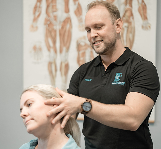
Real-Time Ultrasound imaging (RTUS) provides our team with an accurate and real-time assessment of your pelvic floor muscle strength, control and activation.
Experience Vital Bio-feedback with Real-time Ultrasound Imaging
Increasing your pelvic floor strength can be a long and difficult process. Whether it’s after injury, after prostate surgery or after pregnancy – you may feel lost and uncertain of what you should be doing. It’s hard to know if you’re doing everything correctly when trying to activate your pelvic floor muscles. You might feel like you’re making progress but suddenly hit a plateau.
Archer St Physiotherapy Centre offers external, non-invasive Real-Time Ultrasound imaging to scan your pelvic floor muscles. With this technology, you’ll see what muscles you need to contract, how to contract them, and how strong they are. Seeing this in real-time will help give you a better understanding of the most accurate muscle activations. You can confidently complete the correct exercises and speed up your recovery time with this visual feedback!
Advantages
Advantages Of Real-Time Ultrasound Imaging
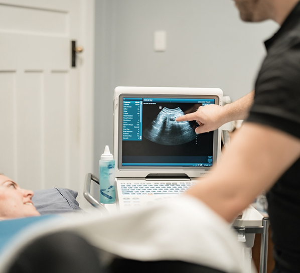
With the help of our state-of-the-art ultrasound scanning machine, patients will be able to watch their core and pelvic floor muscles contract during exercise. This technology makes deep muscle strengthening a more tailored and accurate experience to boost performance levels. Physiotherapists can perform an objective assessment of pelvic and abdominal muscles and their level of muscle function.
Accurate deep muscle activation
The activation of superficial muscles is easily determined through palpation. Ultrasound imaging allows for the observation and assessment of deep core stability and pelvic floor muscles during activation.
Exercise feedback
Feedback during exercise ensures accurate and efficient activation of the necessary pelvic and abdominal muscles, giving the patient confidence and assurance.

Improved recovery time
Increased accuracy of pelvic floor muscle activation results in more efficient retraining of the muscles, leading to increased muscle recovery time.
Knowledge of deep muscle systems
Why Choose Archer St Physiotherapy Centre for Real-Time Ultrasound Imaging

With real-time ultrasound imaging, you’ll have everything you need right in front of you. The physiotherapist will be able to show you on the screen which muscles are working, how to accurately contract them, and how strong they are.
With the help of our state-of-art ultrasound machine, you can see how your deep muscles are contracting during exercise and movement. Physiotherapists can perform an objective assessment of pelvic muscle control and the level of muscle function. Diagnostic ultrasound scanning will allow us to plan and implement an effective home exercise regimen that targets these crucial muscle groups for strengthening, stability and control. Come see us today to learn more about how we can help you achieve your wellness goals!
- Easily Accessible Location in Carlisle with Disability Amenities
- Effective and Hands-On Physiotherapy Every Visit
- Multi-Award Winning Physiotherapy Practice
- Free and Convenient On-site Parking
Frequently Asked Questions
FAQ's about Real-Time Ultrasound Imaging
How does Real-Time Ultrasound Imaging work?
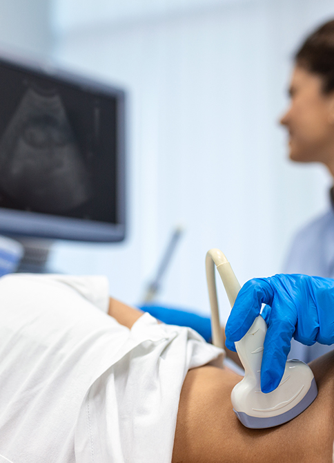
The use of real-time ultrasound imaging is painless, external and non-invasive. A physiotherapist will apply an ultrasound wand to your abdomen to examine the core and pelvic muscles beneath. They will point out the muscle groups on the screen and teach you how to contract them efficiently, using helpful cues. The screen allows you to see the contractions in real-time, which can be helpful in your recovery process.
Therefore, real-time ultrasound imaging can help you achieve better health by showing you how to contract your core and pelvic floor muscles accurately.
How can Real-Time Ultrasound Imaging help me?
Real-time ultrasound has many applications. Physiotherapists commonly use this technology to image muscles that control bladder and bowel continence, with the goal of educating you in how to activate and strengthen these muscles. Weak bladder and bowel control can occur after pregnancy in women, and after prostate surgery or treatment in men. Patients may need assistance with re-strengthening the muscles that control bladder and bowel function, stability and endurance – particularly if they are experiencing light bladder leakage when they sneeze, cough, jump, strain to lift something heavy, or if they are finding it difficult to hold on to go to the toilet.
Assessment of the Core Muscles
Supporting the spine and pelvis requires strong core muscles. It is possible to see the core muscles with ultrasound imaging. We can then identify which layers of muscle are activating when the core is contracted and if there are any problems with accurate contraction.
Real-time or diagnostic ultrasound can also measure abdominal separation. The condition is called rectus diastasis and may occur after pregnancy when the abdominal muscles have been stretched. In addition to using ultrasound for assessment to determine the separation, a physiotherapist can also help you find ways to recover and reduce the separation through exercise prescription.
Evaluation of the Pelvic Floor
Real-time ultrasound imaging technology can also be used to image the pelvic floor and muscles that control continence. Using the images, we can also measure things like bladder function, pelvic muscle strength and control, and help you regain a strong and efficient pelvic floor.